The Cellular Resolution in vivo OCT System
Your Best Partner for Skin Diagnosis
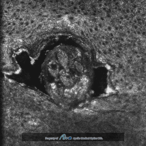
SeeKIN, based on OCT technology, is a skin layer optical scanning device. It provides horizontal and vertical images to observe cellular structure beneath the skin. It also features computation and report generation capabilities.
Our technology can detect and quantify melanosome size, active melanocytes, and collagen healthiness to track and monitor the skin’s condition in response to skincare products.
Embedded video dermatoscope, square color image, 5mm*5mm
Weight
9 Kg
Size
330*351*143 (mm)
Integration of OCT and Image Guiding System
During clinical diagnosis, physicians need to obtain as much information as possible. Therefore, combining the Optical Coherence Tomography (OCT) system with the image guiding function can enhance its clinical usability and is vital to improving examination efficiency.
AMO’s image guiding system allows users to switch between skin image guiding and OCT scanning modes according to different needs. To start off, the user finds the area to be scanned using the image guiding mode. During the scan, the user will switch to the OCT scanning mode, then switch back to the image guidance mode once the scan is complete.
High-speed Dual-mode Scanning Switch
Based on a patient’s various pathologies, detailed images need to be taken in multiple directions in order to provide the physician with sufficient information for interpretation and clinical diagnosis.
The user can easily switch between the cross-sectional scan (B-scan) and the en face scan (E-scan) in just 1 second. This avoids the potentially disastrous issue of overlooking a lesion and makes the process of diagnosis much more efficient.
Three-dimensional Image Stack
AMO’s OCT provides a three-dimensional isotropic resolution up to one micrometer which is essential for a three-dimensional image. ApolloVue S100 could acquire the E-scans continuously and reconstruct the three-dimensional images which allows users to explore the image in any section.
Need help?
Please send your inquiries to our email address and we'll connect you to an expert.


